Home » Uncategories » Shoulder Muscles Diagram / Practice, Practice, Practice, Perform: The Shoulder Is Not ... - It is one of the most mobile joints in the human body, at the cost of joint stability.
Shoulder Muscles Diagram / Practice, Practice, Practice, Perform: The Shoulder Is Not ... - It is one of the most mobile joints in the human body, at the cost of joint stability.
Shoulder Muscles Diagram / Practice, Practice, Practice, Perform: The Shoulder Is Not ... - It is one of the most mobile joints in the human body, at the cost of joint stability.. The following is an overview of the shoulder muscle anatomy. The tendons are the attachment of the muscle to the bone. It is a flat, gliding joint. The shoulder abduction muscles are supraspinatus, deltoid, trapezius, and serratus anterior.a mnemonic memory aid to remember these four muscles responsible for abducting the shoulder is: The rotator cuff is a group of four muscles and tendons that surround the glenohumeral joint.
Postural and active movement muscle, used to tilt and turn the head and neck, shrug, steady the shoulders, and twist the arms. What can you tell us about how these joints work? The shoulder blade (scapula) connects to the collarbone (clavicle) at this joint. It is one of the most mobile joints in the human body, at the cost of joint stability. The muscles of the shoulder support and produce the movements of the shoulder girdle.they attach the appendicular skeleton of the upper limb to the axial skeleton of the trunk.
Stiff Neck? Too Much Office? Let's See How To Release ... from theawesomedaily.com The following is an overview of the shoulder muscle anatomy. The rotator cuff muscles and tendons may be injured by trauma, such as falling when skiing or biking, or from arthritic spurs that form within the shoulder and erode the cuff tissue over time. The partner should slowly, but firmly press on both sides of your shoulder to compress the ac joint. Postural and active movement muscle, used to tilt and turn the head and neck, shrug, steady the shoulders, and twist the arms. The shoulder blade (scapula) connects to the collarbone (clavicle) at this joint. And the ligaments, which connect bones. The tendons are the attachment of the muscle to the bone. The shoulder muscles are responsible for maintaining the widest range of motion of any joint in your body.
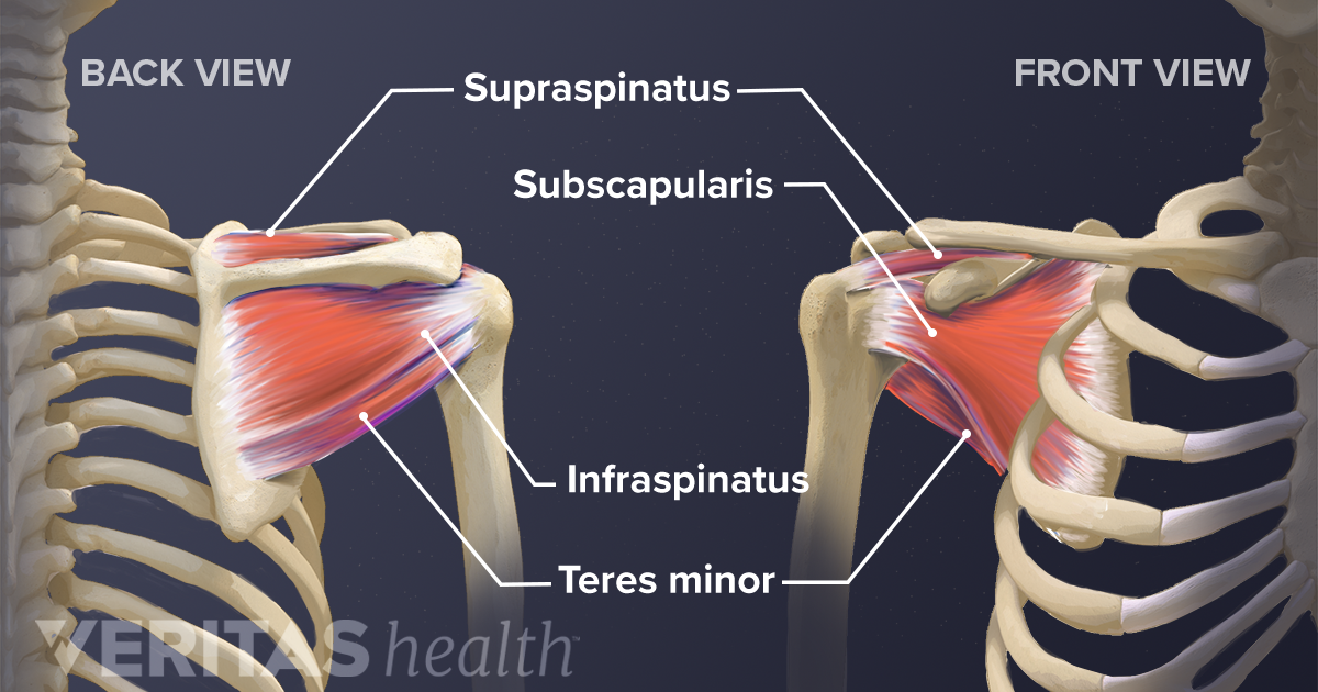
The main shoulder muscles are trapezius, deltoid, pectoralis major and 4 rotator cuff muscles:
To further reinforce the shoulder, the four muscles of the rotator cuff extend from the scapula and surround the head of the humerus to both rotate the arm and prevent dislocation. The following is an overview of the shoulder muscle anatomy. The rotator cuff muscles are important stabilizers and movers of the shoulder joint. The shoulder muscles consist of the deltoids and the rotator cuff group.the deltoids are the muscles that can be seen on the outside of the body, whilst the rotator cuff group is found within the shoulder joint itself, providing structural support and allowing the shoulder to perform many functions. Super dads tickle super alpacas. The most common shoulder injuries are sprains, strains, and tears. This muscle moves each shoulder joint in four distinct ways as well as keeps the arms attached to the body. And the ligaments, which connect bones. A muscle contracts to move bones; The shoulder joint (glenohumeral joint) is a ball and socket joint between the scapula and the humerus.it is the major joint connecting the upper limb to the trunk. The muscles of the shoulder bridge the transitions from the torso into the head/neck area and into the upper extremities of the arms and hands. The shoulder abduction muscles are supraspinatus, deltoid, trapezius, and serratus anterior.a mnemonic memory aid to remember these four muscles responsible for abducting the shoulder is: Muscle structure of the knee 12 photos of the muscle structure of the knee muscle anatomy knee mri, muscle anatomy of the knee, muscle anatomy of the knee joint, muscle and tendon structure of the knee, muscle structure of the human knee, human muscles.
The humeral head in the glenoid socket. Human body anatomy human anatomy and physiology leg muscles anatomy shoulder anatomy muscle diagram dog grooming styles medical anatomy shoulder muscles rotator cuff. A muscle contracts to move bones; And the ligaments, which connect bones. Postural and active movement muscle, used to tilt and turn the head and neck, shrug, steady the shoulders, and twist the arms.
Stretching: How to Stretch the Shoulder | Anatomía ... from i.pinimg.com Around the shoulder, muscles in the back, neck, shoulder, chest and upper arm all work together to support and move the shoulder. What are common rotator cuff injuries? The shoulder girdle is also called the pectoral girdle, and it is a bone ring, incomplete posteriorly.the shoulder girdle is formed by two sets of bones: Muscle structure of the knee 12 photos of the muscle structure of the knee muscle anatomy knee mri, muscle anatomy of the knee, muscle anatomy of the knee joint, muscle and tendon structure of the knee, muscle structure of the human knee, human muscles. Four of them are found on the anterior aspect of the shoulder, whereas the rest are located on the shoulder's posterior aspect and in the back. The most common shoulder injuries are sprains, strains, and tears. Parts of the right shoulder blade: The muscles of the shoulder support and produce the movements of the shoulder girdle.they attach the appendicular skeleton of the upper limb to the axial skeleton of the trunk.
The shoulder abduction muscles are supraspinatus, deltoid, trapezius, and serratus anterior.a mnemonic memory aid to remember these four muscles responsible for abducting the shoulder is:
Muscle structure of the knee 12 photos of the muscle structure of the knee muscle anatomy knee mri, muscle anatomy of the knee, muscle anatomy of the knee joint, muscle and tendon structure of the knee, muscle structure of the human knee, human muscles. The shoulder joint (glenohumeral joint) is a ball and socket joint between the scapula and the humerus.it is the major joint connecting the upper limb to the trunk. Parts of the right shoulder blade: The most common shoulder injuries are sprains, strains, and tears. The shoulder muscles consist of the deltoids and the rotator cuff group.the deltoids are the muscles that can be seen on the outside of the body, whilst the rotator cuff group is found within the shoulder joint itself, providing structural support and allowing the shoulder to perform many functions. Muscles allow us to move by pulling on bones. Related posts of diagram of shoulder muscles and tendons muscle anatomy equine. The shoulder muscles are responsible for maintaining the widest range of motion of any joint in your body. Shoulder muscles move the shoulder blades and upper arm bones. The shoulder blade (scapula) connects to the collarbone (clavicle) at this joint. What are common rotator cuff injuries? Numerous muscles help stabilize the three joints of. The shoulder abduction muscles are supraspinatus, deltoid, trapezius, and serratus anterior.a mnemonic memory aid to remember these four muscles responsible for abducting the shoulder is:
The shoulder's flexibility can make it prone to injury. While seated, have your partner place one hand at the front of your shoulder joint and one hand at the rear. The largest of these shoulder muscles is the. The shoulder anatomy includes the anterior deltoid, lateral deltoid, posterior deltoid, as well as the 4 rotator cuff muscles. It is a flat, gliding joint.
Soft Tissues of the Shoulder from embed.widencdn.net This often happens when stress is placed on the tissues that stabilize the shoulder—the muscles; The rotator cuff muscles and tendons may be injured by trauma, such as falling when skiing or biking, or from arthritic spurs that form within the shoulder and erode the cuff tissue over time. Super dads tickle super alpacas. Superficial muscles are the muscles closest to the skin surface and can usually be seen while a body is performing actions. The scapulae, posteriorly, the clavicles anteriorly and completed anteriorly by the manubrium of the sternum (part of the axial skeleton). It is one of the most mobile joints in the human body, at the cost of joint stability. The rotator cuff is a group of four muscles and tendons that surround the glenohumeral joint. Muscle structure of the knee 12 photos of the muscle structure of the knee muscle anatomy knee mri, muscle anatomy of the knee, muscle anatomy of the knee joint, muscle and tendon structure of the knee, muscle structure of the human knee, human muscles.
The large deltoid muscle is the outer layer of shoulder muscle.
Related posts of shoulder muscles and tendons diagram muscle structure of the knee. This often happens when stress is placed on the tissues that stabilize the shoulder—the muscles; The muscles in the shoulder aid in a wide range of movement and help protect and maintain the main shoulder joint, known as the glenohumeral joint. The most common shoulder injuries are sprains, strains, and tears. A muscle contracts to move bones; Deltoids anatomy when most people think of the Muscle structure of the knee 12 photos of the muscle structure of the knee muscle anatomy knee mri, muscle anatomy of the knee, muscle anatomy of the knee joint, muscle and tendon structure of the knee, muscle structure of the human knee, human muscles. The shoulder girdle is also called the pectoral girdle, and it is a bone ring, incomplete posteriorly.the shoulder girdle is formed by two sets of bones: Parts of the right shoulder blade: While seated, have your partner place one hand at the front of your shoulder joint and one hand at the rear. What are common rotator cuff injuries? The shoulder joint (glenohumeral joint) is a ball and socket joint between the scapula and the humerus.it is the major joint connecting the upper limb to the trunk. Shoulder muscles move the shoulder blades and upper arm bones.
0 Response to "Shoulder Muscles Diagram / Practice, Practice, Practice, Perform: The Shoulder Is Not ... - It is one of the most mobile joints in the human body, at the cost of joint stability."


0 Response to "Shoulder Muscles Diagram / Practice, Practice, Practice, Perform: The Shoulder Is Not ... - It is one of the most mobile joints in the human body, at the cost of joint stability."
Post a Comment